Research Assistant
Stanford Mechanical Engineering
Advisor: Professor Matthias Ihme, Center for Computational Fluids and Turbulence Research
June 2018 - June 2019
Carbon capture and sequestration is a promising method to reduce atmospheric levels of carbon dioxide. CO2 sequestration models can be improved by studying phase changes under nanoconfinement conditions. By replicating behavior in mineral trapping with nanoconfined CO2, we are closer to optimizing sequestration capacities.
Under Prof. Ihme at Stanford, my accomplished objectives were:
1) Prototyping a flow system to examine supercritical carbon dioxide fluid phase transitions under nanoconfinement
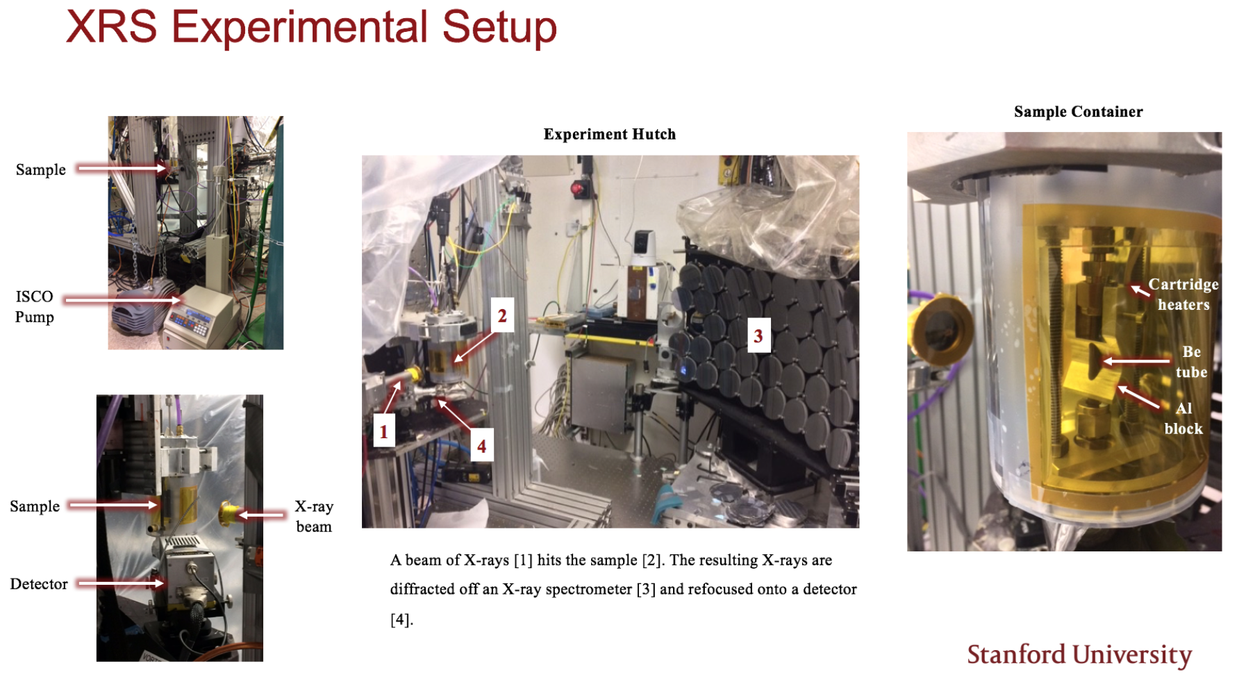
Recent literature has shown simulation evidence of improved carbon capture in porous media at nanoconfinement. I designed and assembled a microfluidics flow system that would allow carbon dioxide to flow at its supercritical state through a chamber of nanoconfined spaces. Beyond selecting tubing and seals to withstand high temperatures and pressures, I researched nanofabrication techniques to achieve materials that could satisfy nanoconfinement conditions and still be imaged using X-ray spectroscopy. The final experimental setup included a carefully selected Teledyne ISCO pump, a custom-made Beryllium imaging chamber, 100nm pellet-pressed silica powder, cartridge heaters, and various steel tubing/Swageloks.
2) Performing X-ray spectroscopy of the nanoconfined fluidics system at SLAC National Accelerator Laboratory
To test the extent of carbon dioxide capture into nanoconfined spaces, I worked closely with Dimosthenis Sokaras for beam access at the SLAC National Accelerator Laboratory. We compared samples of absorption rates of the Beryllium chamber itself, the chamber with pressed silica nanoparticles, and the fully-functioning flow system to determine the presence of supercritical carbon dioxide. The image at the top of this page depicts the main elements of our experimental setup at SLAC.
3) Characterizing nanoconfinement conditions of packed powder using X-ray CT imaging and Matlab
Pellet-pressing the silica powder created nano-sized voids to capture the carbon. However, the true nature and size of these voids need to be imaged and measured with additional X-ray CT imaging. With the X-ray tomography equipment in the Stanford Nano Labs, I took 2D slices of the sample and calculated porosity to characterize the carbon dioxide entrapment.
After completion of my research assistantship with Prof. Ihme, I transferred this project to Priyanka Muhunthan to be completed as her PhD dissertation.
# nanomaterials, microfluidics, X-ray spectroscopy and tomography


Prototype tomography imaging